

If you’ve only ever seen grainy, black-and-white 2D ultrasound images where you need someone to point out which blob is the baby’s head, 3D and 4D ultrasounds are going to blow your mind. Instead of flat, two-dimensional slices, advanced prenatal screening tests create detailed, lifelike images of your baby – and in the case of 4D, you can actually watch them moving in real-time.
But beyond the undeniable “cool factor” of seeing your baby’s face before they’re born, 3D and 4D ultrasounds offer genuine medical benefits. They provide clearer visualisation of fetal anatomy, help detect certain abnormalities that 2D scans might miss, and give parents a deeper connection to their pregnancy.
That said, they’re not necessary for everyone, and standard 2D ultrasounds remain the gold standard for routine prenatal screening. So let’s break down what 3D and 4D scans actually do, when they’re beneficial, and whether they’re worth considering for your pregnancy.
What’s the Difference Between 2D, 3D, and 4D Ultrasounds?
Understanding the technical differences helps clarify what each type of scan offers.
2D ultrasound creates flat, two-dimensional images – cross-sectional views of your baby. These are the standard black-and-white images you see at routine antenatal appointments. They’re excellent for measuring growth, checking organ development, and monitoring pregnancy health.
3D ultrasound uses multiple 2D images taken from different angles and combines them to create a three-dimensional picture. Instead of flat cross-sections, you get detailed surface images showing your baby’s features – their face, hands, feet.
4D ultrasound is essentially 3D in motion – it’s 3D imaging updated in real-time, creating a moving picture. You can watch your baby yawn, suck their thumb, or pull faces. The “fourth dimension” is time.
All three types use the same basic ultrasound technology – sound waves that bounce off tissues to create images. The difference is how the data is processed and displayed.
Medical Benefits of 3D and 4D Ultrasounds
While 2D ultrasounds are sufficient for most prenatal care, 3D and 4D scans offer specific medical advantages in certain situations.
Better visualisation of facial abnormalities: Conditions like cleft lip and palate are much easier to identify and assess with 3D imaging. You can see the entire facial structure rather than trying to interpret it from 2D cross-sections.
Improved detection of skeletal abnormalities: 3D scans provide clearer images of bones and can help identify issues with limb development, spinal problems, or skeletal dysplasias that might be ambiguous on 2D imaging.
More accurate assessment of neural tube defects: While 2D ultrasounds detect most neural tube defects like spina bifida, 3D imaging can provide better detail about the extent and severity of the defect, which helps with treatment planning.
Clearer placental imaging: 3D ultrasound offers better visualisation of the placenta’s position and structure, which is crucial when there are concerns about placenta previa, accreta, or other placental abnormalities.
Enhanced evaluation of fetal abnormalities: When 2D scans show something potentially concerning but unclear, 3D and 4D imaging can provide additional perspectives that help clarify whether there’s actually a problem or just an unusual viewing angle.
Bonding and Emotional Benefits
Beyond the medical advantages, there’s genuine psychological value in 3D and 4D ultrasounds, particularly for parents.
Seeing your baby’s face, watching them move, and getting a clear picture of what they look like creates a stronger emotional connection. For some parents, this makes the pregnancy feel more real and helps them bond with the baby before birth.
This is particularly meaningful for partners who aren’t carrying the baby. They can’t feel the kicks and movements the way the pregnant person can, so seeing detailed images and watching real-time movement helps them feel more involved in the pregnancy.
For families who’ve experienced previous pregnancy loss, 3D and 4D scans can provide reassurance and help them connect with a new pregnancy when anxiety might otherwise dominate.
There’s also something valuable about being able to share clear images with family and friends. Instead of showing people grainy 2D images where they nod politely and pretend they can see what you’re pointing at, you’ve got recognisable pictures of an actual baby.
When Are 3D and 4D Scans Most Useful?
Timing matters enormously for getting good 3D and 4D images. Too early, and the baby hasn’t developed enough detail. Too late, and they’re so squished in there that you can’t get clear facial views.
The sweet spot is usually between 26 and 32 weeks. By this point, your baby has developed fat under their skin, which gives their face definition and makes features visible. They’re also still small enough that there’s amniotic fluid around them, providing clear imaging windows.
Before 24 weeks, babies don’t have enough facial fat to create those adorable, chubby-cheeked images people expect. After 34 weeks, they’re often so cramped and pressed against the uterine wall that getting clear facial views becomes difficult.
If the scan is being done for medical reasons rather than just for fun, the timing might be different depending on what’s being assessed. Your healthcare provider will advise on optimal timing for diagnostic purposes.
Limitations of 3D and 4D Scans
These scans aren’t magic – there are limitations worth understanding before you book one.
Image quality varies: Several factors affect how clear the images are – your body composition, amount of amniotic fluid, baby’s position, placental location. If your baby is facing your spine or has their hands in front of their face, you might not get great views no matter how good the technology is.
They don’t replace standard 2D ultrasounds: 2D ultrasounds are still superior for measuring growth, checking organ function, and routine pregnancy monitoring. 3D and 4D are supplementary, not replacements.
Not all abnormalities are visible: 3D and 4D imaging excels at surface features and skeletal structures, but internal organ problems often require 2D ultrasound for proper assessment.
Requires skilled operators: Getting good 3D and 4D images takes expertise. Not all sonographers are equally experienced with this technology, which affects image quality.
Safety Considerations
Ultrasound technology has been used in pregnancy for decades and is considered safe when used appropriately by trained professionals. There’s no evidence that standard diagnostic ultrasound causes harm to developing babies.
That said, professional medical bodies recommend that ultrasounds should be performed only when medically indicated, by qualified professionals, using the minimum energy necessary to obtain diagnostic information.
Commercial “keepsake” ultrasound boutiques that exist purely for entertainment purposes are controversial. While the technology itself is safe, there are concerns about untrained operators using equipment incorrectly, extended scanning sessions, and the potential for missing medical problems because staff aren’t qualified to recognise them.
If you’re getting a 3D or 4D scan, ensure it’s performed by qualified medical professionals as part of appropriate prenatal care rather than purely as entertainment.
Private vs NHS 3D and 4D Scans
In the UK, routine NHS antenatal ultrasounds use 2D technology. 3D and 4D scans are typically only offered on the NHS when there’s a specific medical indication – when they’ll provide diagnostic information that 2D scans can’t.
If you want a 3D or 4D scan for bonding or keepsake purposes, you’ll usually need to book privately. Many private ultrasound clinics offer 3D and 4D scans as standalone services.
When choosing private scanning services, verify that:
- Sonographers are qualified and registered professionals
- Equipment is properly maintained and calibrated
- There’s appropriate medical oversight
- They have protocols for what to do if they spot potential problems
Our approach to pregnancy health monitoring combines advanced imaging technology with comprehensive medical assessment.
What to Expect During a 3D or 4D Scan
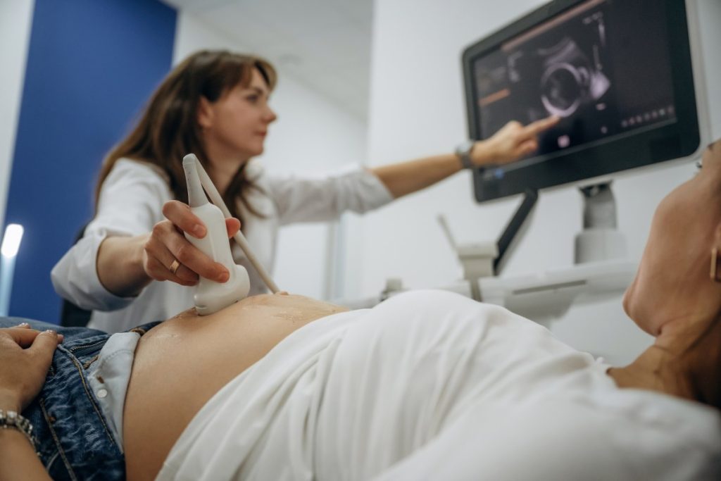
The actual scanning process is similar to standard 2D ultrasounds. You’ll lie on an examination table, gel is applied to your abdomen, and the sonographer moves the transducer over your belly.
For 3D scans, the sonographer captures multiple 2D images from different angles, which are then processed to create three-dimensional pictures. For 4D, you’ll watch real-time moving images on a screen while the sonographer works.
Sessions typically last 20-45 minutes, depending on what’s being assessed and how cooperative your baby is. If the baby isn’t in a good position, you might need to move around, go for a walk, have a snack, or even reschedule for another day.
Most clinics provide photos and sometimes video recordings of the scan. Some offer packages with printed images, USB drives, or online galleries.
Cost Considerations
Private 3D and 4D scans range from around £80 to £200+, depending on the clinic, session length, and what’s included. Packages with longer scanning time, multiple printed images, or video footage cost more.
If you’re having the scan for medical reasons as part of specialist prenatal care, it might be covered by private medical insurance, but purely elective “bonding” scans typically aren’t.
The Bottom Line
3D and 4D ultrasounds offer clear medical benefits for detecting and assessing certain fetal abnormalities, particularly facial, skeletal, and surface conditions. They provide better visualisation than 2D scans in specific circumstances and complement standard prenatal screening.
Beyond medical uses, they offer genuine emotional benefits – helping parents bond with their baby, involving partners more fully in the pregnancy, and providing reassurance and connection.
But they’re not essential for routine prenatal care. Standard 2D ultrasounds remain the gold standard for monitoring pregnancy health and development. 3D and 4D scans are valuable additions when there’s medical indication or when parents want enhanced bonding opportunities, but they’re supplements rather than necessities.
If you’re considering a 3D or 4D scan, ensure it’s performed by qualified professionals as part of comprehensive prenatal care rather than as isolated entertainment. The best outcomes come from integrating advanced imaging with thorough medical assessment and appropriate follow-up care.