
The placenta is one of those things that most people don’t think much about until they’re pregnant, at which point it suddenly becomes very significant indeed. Formed early in pregnancy and attached to the uterine wall, the placenta is responsible for supplying your baby with oxygen and nutrients, removing waste products, and producing hormones that support the pregnancy. It does a remarkable amount of work quietly and without fanfare. But where it implants in the uterus matters, and the position of your placenta is something that will be assessed at your scans and discussed with you if anything requires monitoring.
Where Does the Placenta Usually Sit?
The placenta can implant anywhere on the inner surface of the uterus, and to a large extent its location is a matter of chance. Common positions include the front wall of the uterus (anterior), the back wall (posterior), the top (fundal), or either side. An anterior placenta – sitting towards the front – is very common and generally carries no complications; one thing women often notice is that it can act as a kind of cushion between the baby and the abdominal wall, which means they feel foetal movements later or less intensely than they might otherwise.
A posterior placenta, sitting towards the back of the uterus closer to the spine, is also entirely normal and often allows for earlier, clearer perception of foetal movement.
When Position Becomes Clinically Relevant: Placenta Praevia
The situation that clinicians monitor most carefully is when the placenta implants in the lower part of the uterus, near or over the cervix. This is called placenta praevia, and its significance is related to delivery. If the placenta partially or fully covers the cervical opening, a vaginal birth is not possible, and the way in which labour is managed must be adjusted accordingly.
There are degrees of placenta praevia: a marginal praevia means the placenta reaches close to but not over the cervical os, a partial praevia means it covers part of it, and a complete (or major) praevia means it covers the cervix entirely.
Here’s something important to know, though: a low-lying placenta detected at the 20-week anomaly scan is not necessarily cause for alarm. In the second trimester, a significant proportion of low-lying placentas move upward as the uterus grows and the lower segment expands – a process sometimes referred to as placental migration, though the placenta itself isn’t moving so much as the uterine wall beneath it is stretching. Because of this, women found to have a low-lying placenta at 20 weeks are usually offered a follow-up scan at around 32-34 weeks to reassess position. Many of those placentas will have moved well clear of the cervix by that point.
Major placenta praevia, where the placenta is clearly covering the cervical os at a late gestation, requires careful management. It can cause painless vaginal bleeding in the second and third trimesters, and a planned caesarean section is typically recommended. Women with this diagnosis are monitored closely and advised to seek immediate assessment if any bleeding occurs.
Placenta Accreta Spectrum
Related to, but distinct from, placenta praevia is a group of conditions known collectively as placenta accreta spectrum. These involve abnormal attachment of the placenta to the uterine wall, where the placental tissue grows too deeply into or through the uterine muscle. This is associated with previous uterine surgery, including caesarean sections – the scar tissue from a prior caesarean can disrupt the normal barrier between placenta and uterine wall.
Placenta accreta is rare, but it’s worth awareness because it carries risks related to delivery and postpartum haemorrhage. It is typically identified prenatally through careful ultrasound assessment, sometimes supplemented by MRI, and delivery is planned with specialist input.
Anterior Placenta and Feeling Movements
To return briefly to a more common and less concerning scenario: if you’ve been told you have an anterior placenta and you’re feeling uncertain about when you’ll start feeling your baby move, it’s worth knowing that this is one of the most frequent questions we hear. There’s no magic number of weeks at which movement should begin, but an anterior placenta can genuinely delay the perception of kicks and rolls. This doesn’t mean something is wrong; it just means there’s a bit more between you and your baby in physical terms. Once movement is established, its pattern is what matters.
Placental Position and Pre-eclampsia
There’s also emerging research suggesting associations between placental position and conditions like pre-eclampsia, though this is a complex area and not one where the picture is yet fully clear. What’s well-established is that abnormal placentation more broadly – including issues with how deeply and effectively the placenta embeds – can affect blood supply to the baby and maternal blood pressure. Women with risk factors for pre-eclampsia are monitored accordingly, and understanding more about managing pre-eclampsia risks is useful context for anyone who falls into this category.
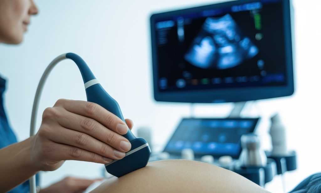
How Position Is Assessed
Placental position is noted at every routine ultrasound scan. At the 12-week dating scan, an early indication of location is often recorded. The 20-week anomaly scan will identify any concern about low-lying position and prompt follow-up if needed. For women with previous caesarean sections, or other risk factors, additional scans may be offered at various stages.
For those who want more detailed assessment or reassurance outside the NHS pathway, private prenatal scan appointments in London are available and can provide specialist sonographer review with time to discuss findings in depth.
The vast majority of pregnancies involve placentas that sit in entirely unremarkable positions and cause no issues whatsoever. But understanding the basics of what clinicians are looking for, and why, means that if anything is flagged at a scan, you’re in a much better position to understand what it means and what happens next.