
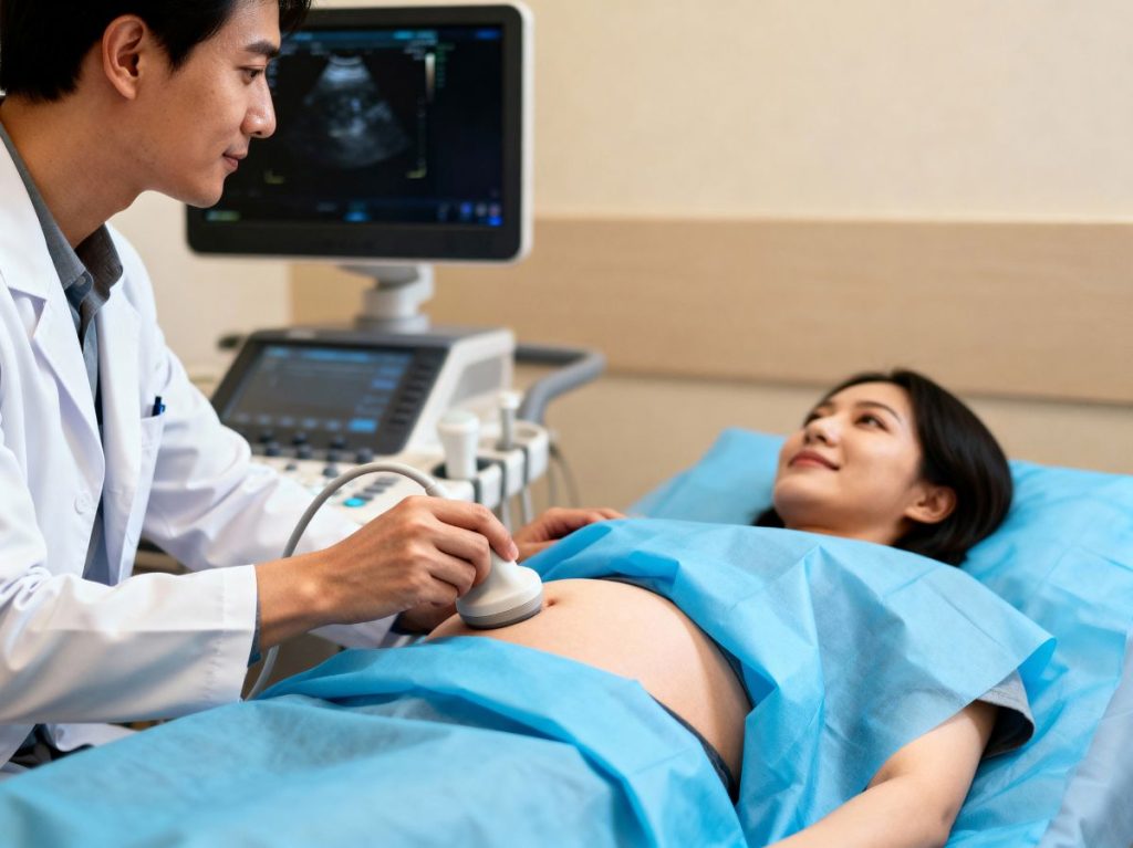
Ultrasound scans are one of those things most pregnant people just accept as part of standard antenatal care without really thinking about why they’re so important. You show up, lie on a table, someone smears gel on your belly and waves a wand around, and you leave with grainy photos of what you’re told is your baby.
But ultrasounds aren’t just for getting those first baby pictures (though that’s admittedly lovely). They’re genuinely crucial diagnostic tools that provide information about your baby’s development, your pregnancy health, and potential complications that can’t be obtained any other way.
Understanding the importance of antenatal scans – what they’re checking for, what they can detect, and how they inform your care – helps you appreciate why they’re standard practice rather than optional extras. Let’s break down the actual importance of ultrasound in pregnancy.
What Ultrasound Actually Does
Ultrasound uses high-frequency sound waves to create images of your baby and reproductive organs. The sound waves bounce off tissues, and the returning echoes are processed to create pictures on a screen.
Unlike X-rays, ultrasound doesn’t use radiation, which makes it safe for frequent use during pregnancy. The technology has been used in obstetrics for decades, and there’s no evidence of harm to developing babies when used appropriately.
What makes ultrasound so valuable is that it provides real-time visual information about things that would otherwise be invisible – your baby’s position, size, movement, organ development, the placenta’s location, amniotic fluid levels. All crucial information for managing pregnancy safely.
Dating and Confirming Pregnancy
One of the first uses of ultrasound in pregnancy is confirming that there’s actually a viable pregnancy and accurately dating it.
Early ultrasounds (usually around 8-14 weeks) can confirm:
- That the pregnancy is in the uterus (not ectopic)
- Whether there’s one baby or multiples
- That the baby has a heartbeat
- How far along the pregnancy is
Accurate dating matters enormously. Your due date determines when screening tests are performed, when certain complications become concerning, and when intervention might be necessary. Calculating dates based on last menstrual period can be quite inaccurate – cycles vary, ovulation timing varies, and not everyone has regular periods.
Early ultrasound dating (ideally between 11-14 weeks) is accurate to within a few days and becomes the official due date for your pregnancy. This precision is crucial for everything that follows.
Detecting Structural Abnormalities
The anomaly scan (usually performed around 20 weeks) is one of the most important ultrasounds of pregnancy. It’s a detailed examination that checks your baby’s anatomy for structural abnormalities or developmental issues.
The scan assesses:
- Brain and skull development
- Face (checking for cleft lip/palate)
- Spine (looking for neural tube defects like spina bifida)
- Heart structure and function
- Abdominal organs (stomach, kidneys, bladder, bowel)
- Limbs (checking all bones are present and properly formed)
- Placental position
- Amniotic fluid levels
Many serious conditions can be detected at this scan – heart defects, kidney problems, skeletal abnormalities, neural tube defects. Early detection allows for planning. Some conditions require specialist care at birth, surgery shortly after delivery, or ongoing management. Knowing about them in advance means the right medical team can be present at birth and treatment can start immediately.
For conditions incompatible with life, early detection gives parents time to make informed decisions about continuing the pregnancy and to prepare emotionally.
Monitoring Fetal Growth
Growth scans track whether your baby is growing appropriately. Babies who are too small (fetal growth restriction) or too large (macrosomia) face different risks and might need closer monitoring or earlier delivery.
Growth restriction can indicate problems with the placenta, infections, chromosomal abnormalities, or other complications. Catching it early means you can be monitored more closely and delivery can be timed to minimise risks.
Larger-than-expected babies might indicate gestational diabetes or other conditions. They’re also at higher risk of birth injuries and might be better delivered via caesarean section.
Without ultrasound, assessing fetal size relies on measuring your bump, which is notoriously inaccurate. Ultrasound provides precise measurements and can track growth trends over time.
Checking Placental Position and Function
The placenta’s position matters enormously. Placenta previa (where the placenta covers the cervix) makes vaginal delivery dangerous and requires caesarean section. Low-lying placenta might move up as pregnancy progresses, but it needs monitoring.
Placental abruption (where the placenta detaches from the uterine wall) is a medical emergency. While ultrasound can’t predict abruption, it can identify risk factors and monitor placental health.
The placenta’s structure and blood flow can also be assessed with ultrasound. Problems with placental function can lead to fetal growth restriction and other complications that require intervention.
Assessing Amniotic Fluid Levels
Amniotic fluid cushions your baby, allows movement necessary for development, and supports lung growth. Too much fluid (polyhydramnios) or too little (oligohydramnios) both signal potential problems.
Low fluid levels might indicate:
- Placental insufficiency
- Fetal kidney problems
- Ruptured membranes
- Post-term pregnancy
High fluid levels might suggest:
- Gestational diabetes
- Fetal swallowing problems
- Twin-to-twin transfusion syndrome (in twin pregnancies)
- Genetic conditions
Ultrasound provides objective measurement of amniotic fluid, which guides management decisions. Severe oligohydramnios might necessitate early delivery; polyhydramnios requires investigation to identify the underlying cause.
Multiple Pregnancy Management
For women carrying twins, triplets, or more, regular ultrasounds are absolutely essential.
Multiple pregnancies need monitoring for:
- Chorionicity (whether babies share a placenta) – this is determined in early pregnancy and affects risk levels and monitoring frequency
- Twin-to-twin transfusion syndrome in identical twins sharing a placenta
- Growth discordance (one twin growing significantly slower than the other)
- Position for delivery planning
Complications in multiple pregnancies can develop rapidly. Regular ultrasound monitoring allows early detection and intervention when needed.
Guiding Procedures
When procedures are necessary during pregnancy – amniocentesis, chorionic villus sampling, fetal blood sampling – ultrasound guidance ensures they’re performed safely and accurately.
Real-time ultrasound imaging lets doctors see exactly where the needle is going, avoiding the placenta and baby while accessing the target area. This significantly reduces risks and improves success rates for these procedures.
Checking Baby’s Position Before Birth
Late pregnancy ultrasounds confirm your baby’s position – head down (cephalic), breech (bottom or feet first), or transverse (sideways).
Position affects delivery planning. Breech babies might be delivered by caesarean section or external cephalic version (manually turning the baby) might be attempted. Transverse babies can’t be born vaginally.
Knowing position in advance prevents surprises during labour and allows time for discussion and planning around delivery options.
Doppler Studies for High-Risk Pregnancies
Doppler ultrasound assesses blood flow through vessels – in the umbilical cord, the baby’s brain, and the uterine arteries. This information is crucial for high-risk pregnancies.
Poor blood flow indicates placental insufficiency or fetal compromise. Monitoring these changes helps determine optimal delivery timing – balancing the risks of prematurity against the risks of continuing pregnancy when the baby isn’t thriving.
Doppler studies are used in pregnancies complicated by:
- Pre-eclampsia
- Fetal growth restriction
- Maternal health conditions affecting placental function
- Twin pregnancy complications
Reassurance and Bonding
While not the primary medical purpose, ultrasounds provide genuine reassurance and help parents bond with their baby. Seeing the heartbeat, watching movement, and getting to “meet” your baby before birth is emotionally significant.
For high-risk pregnancies or parents who’ve experienced previous loss, regular ultrasounds provide ongoing reassurance that the pregnancy is progressing well.
The bonding aspect is particularly valuable for partners who aren’t carrying the baby. Scans help make the pregnancy feel real and tangible for them in ways it already does for the pregnant person.
What Ultrasound Can’t Do
It’s worth noting the limitations. Ultrasound can’t detect all abnormalities – some conditions only become apparent after birth. Image quality depends on factors like maternal body composition, baby’s position, and gestation.
Ultrasound also can’t assess chromosomal conditions like Down syndrome directly – these require blood tests or invasive testing with genetic analysis. Soft markers might raise suspicion, but definitive diagnosis needs other methods.
How Often Do You Need Ultrasounds?
Standard UK antenatal care includes:
- Early scan (8-14 weeks) for dating and viability
- Anomaly scan (18-21 weeks) for detailed anatomy check
High-risk pregnancies require additional scans – growth monitoring, Doppler studies, fluid checks, or other assessments depending on specific concerns.
Low-risk pregnancies might need no additional scans beyond the standard two, though many women choose to have private scans for reassurance or bonding. Prenatal imaging helps track baby development throughout pregnancy while monitoring maternal health and identifying potential complications.
The Bottom Line
Ultrasound is key in modern pregnancy care because it provides information about fetal development, pregnancy health, and potential complications that can’t be obtained any other way.
From confirming pregnancy and accurate dating to detecting abnormalities, monitoring growth, assessing placental function, and checking fluid levels – ultrasounds guide decision-making throughout pregnancy and help ensure the best possible outcomes for both mother and baby.
While the photos are a lovely bonus, the real importance of ultrasound lies in the diagnostic information it provides and how that information shapes your antenatal care and delivery planning. It’s not just about seeing your baby – it’s about keeping both of you safe and healthy throughout pregnancy.